Request an Appointment or call us
What is an Acetabular Fracture?
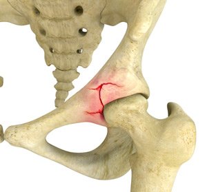
An acetabular fracture is a break in the acetabulum (ball-and-socket portion) of the hip joint. It usually occurs during high-energy injuries.
What are the Causes of Acetabular Fractures?
Causes of acetabular fractures include:
- Automobile accidents
- Osteoporosis
- Falls from heights
- Weak bones
What are the Symptoms of Acetabular Fracture?
Symptoms of an acetabular fracture include:
- Pain
- Hematoma
- Weakness due to nerve damage
- Swelling
- Tingling sensation
- Bruising
How is Acetabular Fracture Diagnosed?
Your doctor will review your medical history and symptoms and a physical examination of the hip will be performed. Your doctor may recommend the following diagnostic tests:
- X-ray: This study uses high-energy electromagnetic energy beams that produce images of the bones.
- CT scan: Special x-rays are used to produce images of any damage to the hip.
- MRI Scan: An imaging study that uses a large magnetic field and radio waves to detect any damage to the soft tissue.
- Bone scan: A nuclear imaging study that helps your doctor to detect any hidden stress fractures or bone disorders.
What is the Treatment for an Acetabular Fracture?
Treatment for acetabular fracture includes:
Non-Surgical Method
- Medications: Your doctor will recommend over-the-counter pain medications to reduce inflammation and pain.
- Physical therapy: Your doctor will recommend special exercises and other techniques to strengthen the bones and muscles.
- Positioning aids: You will be advised to use a leg positioning device such as a knee immobilizer or abduction pillow to help support the restricted leg.
Surgical Method
If non-surgical methods fail to improve the symptoms, surgery will be recommended based on the severity of the fracture. Surgical treatments can include:
- Total Hip Replacement: The damaged bone and cartilage are removed from the hip joint and replaced with artificial components.
- Open Reduction and Internal Fixation (ORIF): The fracture site is exposed, and internal fixation is performed with wires, screws, and nails that are attached to a metal plate placed inside the body.
- Traction: This method involves using a force to gently pull on the injured area to guide the fractured bone ends to their correction position. There are two methods of traction:
- Skin traction: Skin traction involves the attachment of traction tapes to the skin of the hip segment near the fracture.
- Skeletal traction: In skeletal traction, a pin is inserted through the bone and attached to ropes. Weights are applied, and you will be placed in a traction apparatus.
For more information about our services Hip, Knee or to schedule an appointment, call us at or click here to request an appointment online. We’ll respond to you as soon as possible.